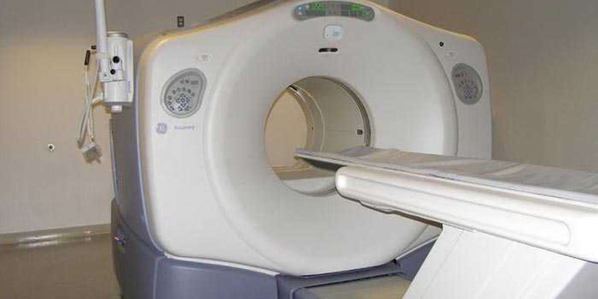

Advanced Diagnostic Imaging: PET/CT Scanning in Alamogordo
What is PET/ CT Imaging Alamgordo?
CT scanning, on the other hand, uses X-rays to create detailed images of the inside of the body. The X-ray machine rotates around the body, taking images from different angles. The images are then combined to create a 3D image of the body, allowing doctors to see detailed structures and identify any abnormalities.
When PET and CT scans are combined, they provide a more complete picture of the body. The PET scan provides information about metabolic activity, while the CT scan provides detailed anatomical information. This can help doctors identify and locate abnormalities in the body, such as cancerous tumors, infections, or inflammation.
Why is PET/CT Imaging Important?
PET/CT imaging is an important diagnostic tool for many different conditions. It is particularly useful in diagnosing and monitoring cancer, as cancer cells often have higher metabolic activity than normal cells. By injecting a tracer that is taken up by cancer cells, doctors can identify the location and extent of the cancer, as well as monitor the effectiveness of treatment.
PET/CT imaging is also useful in diagnosing and monitoring other conditions, such as heart disease and neurological disorders. By providing detailed images of the inside of the body, doctors can identify areas of inflammation, blockages, or other abnormalities that may be causing symptoms.
PET/CT Imaging in Alamogordo:
In Alamogordo, PET/CT scanning is available at several medical centers, including Gerald Champion Regional Medical Center and New Mexico Oncology Hematology Consultants. These centers provide advanced diagnostic capabilities for patients in the area, allowing doctors to identify and treat a wide range of conditions.
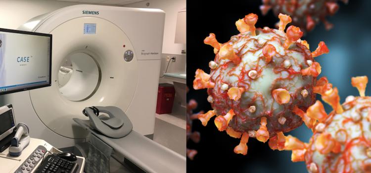
Gerald Champion Regional Medical Center is a 99-bed acute care hospital that provides a wide range of medical services, including PET/CT scanning. The hospital's imaging center is accredited by the American College of Radiology, ensuring that patients receive high-quality diagnostic imaging services. The PET/CT scanner at Gerald Champion Regional Medical Center uses advanced technology to provide detailed images of the inside of the body, allowing doctors to diagnose and monitor a wide range of conditions.
New Mexico Oncology Hematology Consultants is a medical practice that specializes in the treatment of cancer and blood disorders. The practice has several locations throughout New Mexico, including one in Alamogordo that provides PET/CT scanning services. The practice's PET/CT scanner is accredited by the Intersocietal Accreditation Commission, ensuring that patients receive high-quality imaging services. The practice's PET/CT scanner uses advanced technology to provide detailed images of the inside of the body, allowing doctors to diagnose and monitor cancer and other conditions
PET/ CT Imaging Alamgordo How Its Work?
PET/CT imaging is a powerful diagnostic tool that combines two different types of imaging technologies to provide a detailed view of the inside of the body. The combination of Positron Emission Tomography (PET) and Computed Tomography (CT) scans allows doctors to identify and locate abnormalities in the body, including cancer, infections, and heart disease. Here's how PET/CT imaging works:
- The Patient Receives a Tracer:
To begin the PET/CT imaging process, the patient receives a small amount of a radioactive substance called a tracer. The tracer is injected into the patient's body, typically through a vein in the arm. The tracer is chosen based on the condition being diagnosed or monitored, and is designed to accumulate in areas of the body with high metabolic activity.
- The Tracer Emits Positrons:
As the tracer moves through the body, it emits positrons, which are a type of subatomic particle. The positrons interact with electrons in the body, producing gamma rays.
- The Gamma Rays Are Detected:
The gamma rays produced by the tracer are detected by the PET scanner, which is a large, ring-shaped machine that surrounds the patient. The PET scanner detects the gamma rays and creates an image of the body that shows areas where the tracer has accumulated. This allows doctors to see areas of high metabolic activity in the body.
- The CT Scanner Provides Detailed Anatomical Information:
At the same time as the PET scan is being performed, the patient is also undergoing a CT scan. The CT scanner uses X-rays to create detailed images of the inside of the body. The X-ray machine rotates around the patient, taking images from different angles. The images are then combined to create a 3D image of the body.
- The Images Are Combined:
The images from the PET and CT scans are then combined to create a more complete picture of the inside of the body. The PET scan provides information about metabolic activity, while the CT scan provides detailed anatomical information. By combining the two images, doctors can identify and locate abnormalities in the body, such as cancerous tumors, infections, or inflammation.
- The Images Are Interpreted by a Radiologist:
Once the images have been created, they are interpreted by a radiologist, who is a medical doctor with specialized training in diagnosing and treating conditions using medical imaging. The radiologist will review the images and provide a report to the patient's doctor, who will use the information to make a diagnosis and develop a treatment plan.
If you want to get amazing benefits by using this link
Conclusion:
In conclusion, PET/CT imaging is a powerful diagnostic tool that provides detailed information about the inside of the body. By combining PET and CT scans, doctors can identify and locate abnormalities in the body, including cancer, infections, and heart disease. This allows for earlier and more accurate diagnosis, and can improve the outcomes for patients with a wide range of conditions.